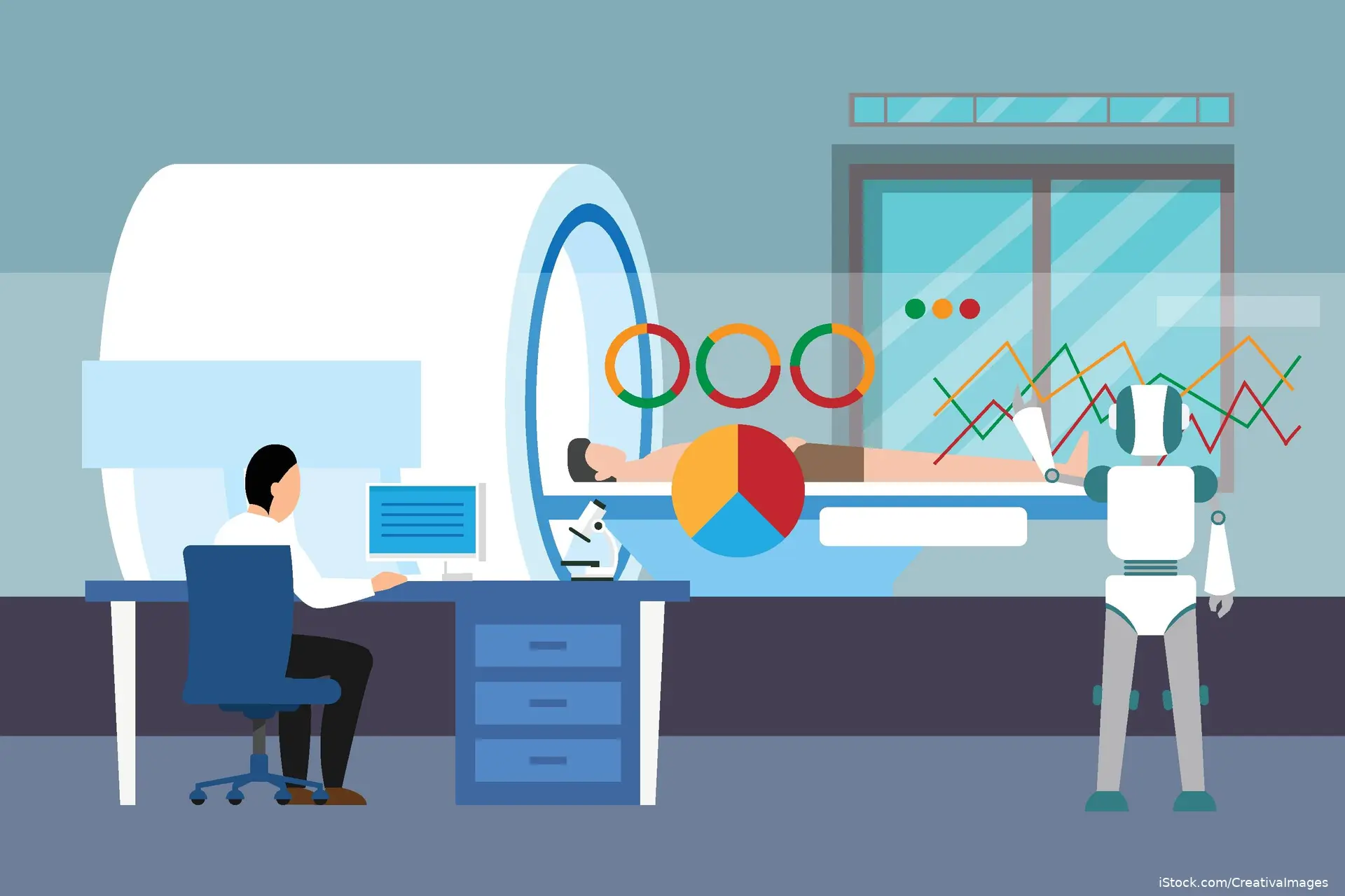
An artificial intelligence system used as an additional reader of contrast-enhanced computed tomography scans helped clinicians identify overlooked liver lesions and prompted changes in radiology reports and clinical management during routine deployment, according to findings published in Nature Medicine.
The Liver DiagnOsis Network, or LiON, was trained on 6,443 patients and retrospectively validated across 22,251 patients from multicenter and real-world cohorts. The system integrates multiphase imaging and clinical information to support liver malignancy diagnosis within existing radiology workflows.
LiON achieved an area under the receiver operating characteristic curve of 0.975 in the retrospective validation. Performance remained high among patients with hepatic steatosis and cirrhosis, with AUCs of 0.971 and 0.924, respectively.
Investigators then evaluated LiON as an additional reader in a single-arm clinical trial involving 10,333 patients in routine clinical practice. The system achieved an AUC of 0.952 and met the prespecified primary performance endpoint. AI-human collaboration identified 51 previously overlooked lesions, including 15 malignancies, and prompted 37 amended radiology reports, 22 multidisciplinary team escalations, and clinical management changes in a subset of patients.
The findings suggest that workflow-integrated AI may serve as a diagnostic safety net alongside radiologists. Because the trial did not include a concurrent comparator group, prospective comparative studies across diverse healthcare systems are needed to determine whether this approach improves clinical outcomes.
Source: Zhang X, Li C, Han X, et al. Large-scale AI-guided liver malignancy diagnosis: multicenter study and a single-arm trial. Nat Med. Published online August 19, 2026. doi:10.1038/s41591-026-04589-y


Advertisement
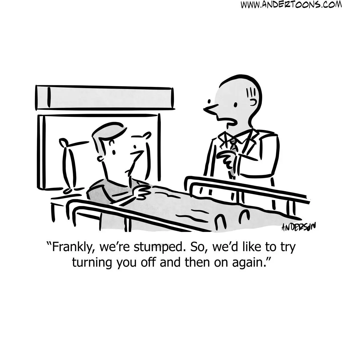
Today's Joke
Cartoon Slide Show

Quiz

Medical Trivia 9/9/2026
1 questions · ≈ 1 min
to compete in the ranking.
×
Loading